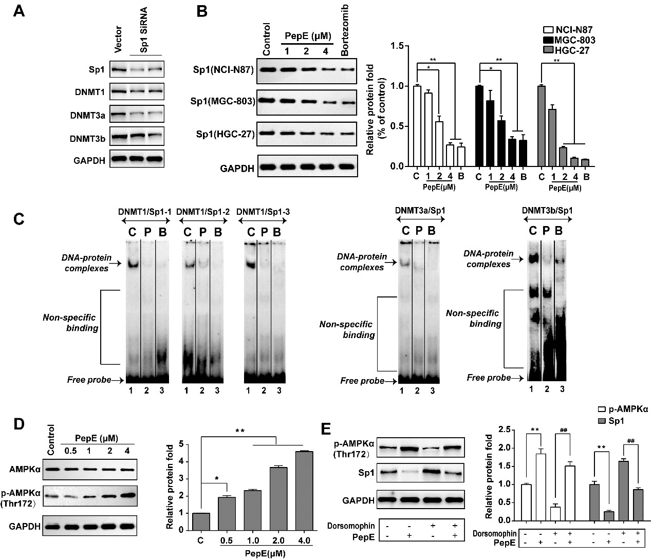
Fig. 8. PepE inhibits AMPKa-Sp1-DNMT signaling in GC cells. (A) Sp1 siRNA concurrently decreased Sp1 and DNMTs (including DNMT1, 3a and 3b) expression in NCI-N87 cells. NCI-N87 cells were transfected with negative control SiRNA (vector) or Sp1 siRNA (1 or 2) and cultured for 2 weeks. Total cell lysates were subjected to Western blot for Sp1, DNMT1, 3a and 3b. (B) PepE inhibits Sp1 protein expression in GC cells. Reduced Sp1 expression in NCI-N87, MGC-803 and HGC-27 cells treated with indicated dosage of PepE for 24 h. Bortezomib was used as positive control (C:control, B:Bortezomib, 100 nM). Data are presented as mean ± SD (n=3), *P<0.05; **P<0.01. (C) PepE abolished Sp1 binding to the DNMT1, 3a and 3b promoter. EMSA was performed with nuclear extracts prepared from NCI-N87 cells untreated or treated with PepE or bortezomib. The DNMT1/Sp1, DNMT3a/Sp1 and DNMT3b/Sp1 probes are shown on the top of each panel. C indicates control; P: PepE (2 µM), and B: bortezomib (100 nM). (D) PepE induced phosphorylation of AMPKa in NCI-N87 cells at indicated doses for 24 h. Data are presented as mean ± SD (n=3). *P<0.05; **P<0.01 when compared with control. (E) The inhibition ability of PepE against Sp1 expression was abrogated by dorsomophin (an AMPK inhibitor). NCI-N87 cells were treated with dorsomophin (10 µM) for 2 h before exposure of the cells to PepE (4 µM) for an additional up to 24 h. Afterward, the expression of p-AMPKa and Sp1 were detected by Western blot. Data are presented as mean ± SD (n=3). **P<0.01 when compared with control, ##P<0.01 when compared with NCI-N87 cells only treated with dorsomophin.